Friday, February 18, 2011

Cone Beam Computed Tomography (CBCT): A Proper Introduction

At Superstition Springs Endodontics (SSE), we have recently integrated Cone Beam Computed Tomography (CBCT) into our practice of endodontics . In my opinion, CBCT is the future of endodontics and the applications in endodontics seem almost limitless. For this reason, we have chosen to implement this technology into our practice.

However, here are a few basic principles about CBCT that might be helpful.
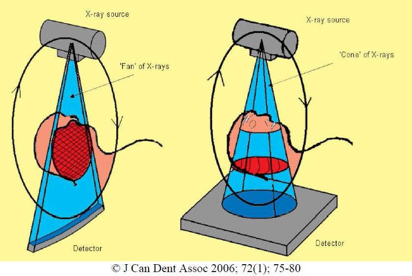
Traditional Computed Tomography (CT or CAT Scan), uses a linear detector and a fan shaped X-ray beam (diagram on left). The detector spins around the patient multiple times over the area to be scan. The computer then takes all of these slices and puts them back together into a 3D image. CT scans are very effective at differentiating between soft and hard tissues.

With Cone Beam Computed Tomography (CBCT), a large detector is used, a cone shaped X ray beam is used, and all data is gathered in a single pass (diagram on right).

This makes the CBCT a smaller unit with the ability to focus the radiographic exam, a single pass around the patient, less radiation exposure and less costly procedure.
CBCT is most effective at evaluation of hard tissues. An important fact to point out is that radiation exposure with CBCT in general is significantly less than traditional CT. Some CBCT units have reduced the radiation exposed to a fraction of the amount of traditional CT.

The field of view (FOV) is an important factor in selecting a CBCT. CBCT has the ability to take a large FOV (full skull), medium FOV (Mx & Md) or focused field (quadrant).

A voxel is a 3 dimensional pixel. The resolution of a CBCT image is determined by the voxel size, as well as the unit's ability to gather & interpret the data with minimal interference/noise.

CBCT units with smaller voxel size are more effective for applications in endodontics, allowing us to see the detail needed.


There have been concerns about the additional radiation exposure associated with CBCT. For the reasons described above, the focus field CBCT has a fraction of the radiation exposure associated with traditional CT. Here are the numbers on radiation...

Since radiation affects different body tissues differently, an effective dose compares the radiation dosage on different body parts on an equivilant basis. The unit for effective dose is the sievert (Sv) joules/kilogram.

In medical procedures, the millisievert (mSv) is used to measure the effective dose.

We are all exposed to about 3.0 mSv/year of natural radiation. Natural radiation comes from the earth as well as from the atmosphere/space.

The following table shows the radiation exposure from several dental radiographic sources as well as medical CT and environmental exposure.

An average American receives 0.0082mSv per day in normal background radiation.

As you can see, one periapical film with d-speed film exposes a patient to 0.0095mSv (which is the equal to about 1.1 days of natural radiation).

A digital periapical film exposes a patient to 1/3 the amount of radiation, 0.0032mSv (which is the equal to about 0.36 days of natural radiation).

The radiation exposure of several CBCT units is listed below.

The amount of radiation exposure is minimal. As with any diagnostic radiographic image, we must balance the need for the information vs. the potential harmful effects of radiation.

The radiation level associated with a CBCT at SSE is the lowest radiation level of any dental CBCT currently available on the market.

Some have inquired into which CBCT that we are using at Superstition Springs Endodontics. We selected the Veraviewepocs 3De by J. Morita. The features we like about it are its high resolution, fast speed, panorex feature, low radiation level and ease of use.

Since many of you will not be able to visit our office to see this in action, the following video clip shows the ease of use and speed of our CBCT.


The following video clip also shows some of the additional benefits of CBCT.


If you are in the Phoenix east valley, we would invite you to contact Annette at our office to schedule a "lunch & learn" and CBCT demonstration (annette@superendo or 480 807-8022). This would include a live demonstration and introduction on how to read a CBCT scan.

If you are in the Phoenix area and would like more information regarding J. Morita or any other CBCT systems (Sirona, Instrumentarium, Soradex, Planmeca or NewTom), contact Jordan at (jordanlhales@gmail.com or 480 227-0435). He will get you any product information for your review.

Tuesday, February 1, 2011

Pulpal Regeneration - More Evidence

In previous posts regarding pulpal regeneration, we have shown not only apical closure, but some dentinal bridging coronal to the apex. While difficult to believe, we have also seen teeth become responsive once again to electric pulp testing. The tissue that forms during pulpal regeneration has been described as "pulp-like", which includes some kind of innervation.

The following case was re-evaluated at 1 year using CBCT. The ability to evaluate apical closure in 3 dimensions is a new way of evaluating success of apexification/apexigenesis/regenerative procedures.

The following case was previously reported and includes the CBCT follow up.


Apical closure at 6 months is noted in regular films.

1 yr recall using CBCT not only shows the apical closure, but coronal bridging under the MTA barrier. In this case a collagen plug was placed prior to MTA. This accounts for the space between the MTA and the new dentinal bridging.

A sagittal view of the tooth also shows complete apical closure as well as coronal dentinal bridging. In this particular case, the MTA plug was not placed deep enough below the enamel and staining occurred, despite using white MTA.

The teeth responded to electric pulp testing with the following readings
#7 - 31 & 36
#8 - 42 &38
#9 - 29
#10 - 28

We recently completed internal bleaching. We removed the MTA down to the layer of collagen plug. Staining noted at the WMTA-dentin barrier. We mechanically removed as much of stain as possible, placed a new coronal barrier and then bleached with the Ultradent endodontic bleaching product. The tooth now looks great. (Sorry no photos on this part)

Wednesday, January 19, 2011

CBCT as a Tool in Endodontic Diagnosis

Cone Beam Computed Tomography (CBCT) is a valuable tool in endodontic diagnosis. The following case illustrates how CBCT provides added diagnostic information not available through traditional 2D imaging.

This patient was referred to our office today after a long week of infection and diagnostic dilemmas. Here's the story...

10 days ago with an ear ache.
9 days ago pt reports pain to chewing & closing teeth together.
8 days ago swelling began. Pt went to ER and was given zithromax, ibuprofen & tylenol #3.
7 days ago swelling increased under tongue and into face.
5 days ago, pt returned to ER where they did a CT scan and found nothing. Pt reports numbness in lip. Pt admitted to hospital and given IV clindamycin. MRI done and "something was found in lower left jaw". Pt started 300mg clindamycin.
Today, patient referred from oral surgery for endodontic consult/vitality testing. Here's how he looked.


Radiographs fairly inconclusive. #18, #19, #20, #21 all normal to percussion, probing and thermal testing.

A small crack noted on the distal marginal ridge of #18. Thermal testing once again indicates a vital pulp. Typically, we would expect a necrotic tooth to be the source of the submandibular swelling that this patient has experienced.

Since tooth #18 is responding normally to thermal testing, we decided to take a CBCT to look for more evidence of the source of infection.

This coronal slice (.25mm) shows radiolucency around the distal root #18. This image is more conclusive than the standard 2D image.

A sagittal slice through the distal root of #18 shows the lesion and its perforation of the lingual plate.

An axial view of the distal root of #18 also shows perforation to the lingual.

These CBCT slices are conclusive enough to revise the pulpal diagnosis to "partially necrotic" and recommend endodontic treatment. It appears that the distal root is necrotic and the infection is spreading through the lingual plate.


RCT initiated. Upon access, we find vital pulp tissue in the mesial canals, and necrotic pulp tissue in the distal canal.

Further removal of the distal crack finds the crack extending down the distal root, below the CEJ. Extraction is recommended.

In endodontic diagnostics, we typically classify pulpal status as:

1. Normal
2. Reversibly Inflammed
3. Irreversibly Inflammed
4. Necrotic

However, things are not always a cut an dry as that. This case illustrates that "partially necrotic" pulp is a possible classification of pulpal status.

Following removal of the tooth, the infection quickly resolved.

CBCT is an important tool for diagnostic imaging in endodontics.

Monday, December 27, 2010

CBCT to Evaluate Internal Root Resorption

CBCT can be used to evaluate the extent of root resorption. Before this technology, we would have to excavate a resorptive defect to evaluated the extent and restorability. The CBCT allows us to see in all dimensions the extent of a resorptive defect. In this case, the non-restorability was determined with CBCT alone. This saves the patient and clinician time and money.

This video is made of individuals slices 1.0mm thick with 0.25mm interval between each slice.

Thursday, December 16, 2010

Monday, November 29, 2010

Successful Perforation Repair using MTA

This patient presented for treatment of #30 in March 2009. Prior RCT had been done and a large furcal lesion as well as periapical lesion were noted. Retreatment was recommended. Upon access, we found 2 additional canals as well as a furcal perforation. The tooth was obturated and perforation was repaired using MTA.

Post-Op films shows the MTA repair in the furcal area. Note the large lesion around the mesial root.

At 6 months, furcal and periapical lesion are improving and the tooth is functional.


At 18 months, the lesion continues to improve, tooth is completely asymptomatic and functional. Proper endodontic treatment and repair with MTA has retained a tooth that many would have considered "hopeless" or non-restorable based on the amount of furcal bone loss.

Wednesday, November 17, 2010

Superstition Springs Endodontics goes 3D


Superstition Springs Endodontics is excited to introduce cone beam technology (CBCT) into their practice of endodontics. The decision to incorporate this technology has come after a extended review of the technology, research and clinical applications of CBCT in endodontics.

Dr. Edward Carlson was among the first endodontists in Arizona to incorporate the operating microscope into his practice of endodontics almost twenty years ago. Just as the operating microscope has become an indispensable tool in the practice of endodontics, we expect CBCT to become integral part of endodontic diagnosis, treatment & evaluation.

As Superstition Springs Endodontics, we are specialists in saving teeth. The CBCT is another diagnostic tool to allow us to make important decisions about saving teeth. Doctors and patients who are committed to saving natural teeth, will be able to benefit from this new technology.

The clinical applications of CBCT in endodontics include:
1. Aid in endodontic diagnosis
2. Canal morphology
3. Evaluation of root fracture
4. Evaluation of internal root resorption
5. Evaluation of invasive cervical resorption
6. Presurgical assessment
7. Evaluation of non-endodontic pathology
8. Assist with implant planning for non-restorable teeth

We look forward to sharing cases using this new technology.

The first case to share is the case of a fractured tooth. This patient had a fall and hit her face 5 months ago. #8 was damaged and had to be removed and replaced with an immediate implant. #9 continued to give her symptoms and mobility.

Now it is obvious with a regular radiograph that there is a problem with this root. The tooth exhibited class II mobility.


The coronal view (left) shows the similar view to the standard radiograph, however, the sagittal view (right) shows how the fracture has sheared off toward the palate, well below the level of palatal bone. The ability to see this fracture from the sagittal view allows us to make a determination of the restorability of this tooth.

Previously, we would have had to remove the fractured portion of the tooth and visualize the depth of the fracture. The CBCT allows us to visualize this without the need to disassemble the tooth. This tooth has been recommended for extraction and the CBCT scan can also be used to help in the treatment planning of the new implant.

Stay tuned for more applications of CBCT in our endodontic practice.

We have selected a CBCT manufactured by J. Morita. J. Morita has been a leader in development of cone beam technology. The Veraviewepocs 3De is a focus field cone beam with incredible resolution, ideal for the practice of endodontics.


Wednesday, November 10, 2010

An inexpensive solution for transillumination

At the most recent Inner Space Seminar, we discussed all different kinds of cracks in teeth. We reviewed how to detect them, classify them, treat them, & prevent them. An effective way to identify cracks in the crown of a tooth is by using transillumination.

I mentioned an inexpensive light that can be used for transillumination. Thanks to Dr. Nathan Saydyk for his research, this light has been discontinued and replaced with the new Browning 2120 Microblast Pen Light with Bore Light Adapter. This is a flashlight used for firearm inspection and cleaning that can be used for transillumination.


Monday, November 1, 2010

Managing a Cracked Tooth

Dealing with cracked teeth can be very challenging. In the first place, there is a lot of confusion about what we are calling a cracked tooth. Craze lines, fractured cusps, split teeth and vertical root fractures are all often called "cracked" teeth. However, treatment and prognosis are different for all of these different situations.

Cracks in teeth are findings, not a diagnosis. Proper pulpal and periapical diagnosis as well as the location and extent of a crack are needed to determine a proper treatment plan. The problem with cracks in the tooth are the possibility for future bacterial penetration, which leads to inflammation and disease.

With these considerations, many teeth with cracks can be saved. Keys to saving teeth with cracks are:
1. Early detection and treatment
2. Proper endodontic diagnosis
3. Proper determination of the location and extent of a crack

The following case of a cracked tooth was recently treated at Superstition Springs Endodontics.

This patient presented with mesial decay on #14 causing discomfort. The tooth was normal to percussion, probing and no response to thermal test. DX: Necrotic pulp w/ normal periapex. A crack was noted on the distal marginal ridge. RCT recommended.

Removal of decay and access revealed the crack extending down the distal wall.

Closer examination finds that the crack ends near the level of the CEJ. Pt is informed of the crack and the prognosis is good, since the new crown will be able to cover the crack. The crack should be removed at the time of the build-up.

A main key to saving teeth with cracks is to identify the location and extent of a crack.

An upcoming Inner Space Seminar, entitled "Breakdance" will help clinicians know how to identify and classify cracks in teeth, as well as treatment plan restorative options for teeth with cracks.

Monday, August 30, 2010

Would you implant or do RCT? - UPDATED

I work with some great oral surgeons & periodontists. I was recently asked to evaluate tooth #31 by my periodontist colleague. This patient had been referred to him for extraction and placement of an implant.


The periodontist realized that the bone loss around this root was not caused by periodontal disease. The patient reported no pain or swelling. He has no senstivity to percussion, normal probing depths (4mm depth on the buccal was the deepest) and when proper vitality testing was completed, the tooth was found to be necrotic. The tooth was diagnosed: Necrotic Pulp w/ Chronic Apical Periodontitis. The patient was given the option of endodontic therapy to retain the natural tooth.


Pulpal access revealed a necrotic pulp chamber.


Endodontic therapy completed and a 6 month recall scheduled to evaluate the periapical healing.
Please feel free to share your thoughts about these cases. The purpose of this blog is to generate discussion. What would you have done?


OK, here we are 3 years later. The tooth is asymptomatic and functional and perio probings are normal. Significant healing has occurred. There is still some lateral radiolucency - widened pdl, but at this point I think it was a good decision to retain the tooth.

Our specialty at Superstition Springs Endodontics is saving teeth.